Posts

Advanced imaging tips T cell target recognition on its head

T cells represent a key component of our immune system, and play a critical role in protecting us against harmful pathogens like viruses and bacteria, and cancers. The more we understand about how they recognise, interact with and even kill infected or cancer cells moves us closer to developing therapies and treatments for a range of conditions.

In a paper published today in the premier international journal Science, an Australian team of scientists led by Monash University, the Australian Research Council Centre of Excellence in Advanced Molecular Imaging and the University of Melbourne at the Doherty Institute, has redefined what we thought we knew about T cell recognition for the past 20 years.

In order to interact with other cells in the body, T cells rely on specialised receptors known as T cell receptors that recognise virus or bacteria fragments that are bound to specialised molecules called major histocompatibility complex (MHC) or MHC-like. Over the past 20 years, the prevailing view was that T cell receptor sat atop the MHC and MHC-like molecules for recognition.

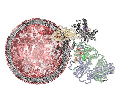
The team of scientists characterised a new population within a poorly understood class of T cells called gamma delta T cells that can recognise an MHC-like molecule known as MR1. Using a high intensity X-ray beam at the Australian Synchrotron, the scientists obtained a detailed 3D image of the interplay between the gamma delta T cell receptor and MR1 revealing an intriguing result whereby the gamma delta T cell receptor bound underneath the MHC-like molecule for recognition. This highly unusual recognition mechanism reshapes our understanding of how T cell receptors can interact with their target molecules, and represents a major development in the field of T cell biology.

“Think of it like a flag attached to a cell. We always thought the T cells were coming along and reading that flag by sitting atop it. We have determined that instead, some T cells can approach and interact with it from underneath,” said Dr Jérôme Le Nours from the Monash Biomedicine Discovery Institute, co-lead author on the paper.

“These are the types of fine and important details that can change how we approach future research avenues in T cell biology,” said Dr Le Nours.

“This is important because T cells are a critical weapon in our immune system, and understanding how they target and interact with cells is crucial to harnessing their power in therapies such as infection and cancer immunotherapy,” he said.

”Our study shows that MR1 is a new type of molecular target for gamma delta T cells. These cells play a decisive role in immunity to infection and cancer, yet what they respond to is poorly understood. MR1 may be signalling to gamma detla T cells that there is a virus, or cancer cell and triggering these cells to initiate a protective immune response,” said Dr Nicholas Gherardin from the Doherty Institute, co-lead author on the paper.

“We’re very excited to follow up these findings in studies that will aim to harness this new biology in disease settings,” he said.

The research findings were a culmination of a six-year project that involved collaborative support from Australian scientists, the ARC Centre of Excellence in Advanced Molecular Imaging, the use of the Australian Synchrotron, and funding from the National Health and Medical Research Council and the Australian Research Council.

Read the full paper in Science titled A class of γδ T cell receptors recognize the underside of the antigen-presenting molecule MR1.

Image: Artwork of a gamma delta T cell receptor interacting with MR1.  Artwork by Dr Erica Tandori.

 

Original article

Opening the world of cancer research to the low vision community

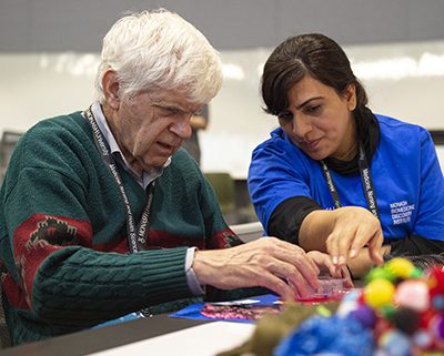
The Monash Biomedicine Discovery Institute (BDI) in collaboration with the Australian Research Council Centre of Excellence in Advanced Molecular Imaging recently opened its doors to the blind and low vision community with the Sensory Scientific Exhibition and Discovery Day. Held on Friday 6 December, also including students from local primary and secondary schools, and members of the wider community, more than 70 attendees had the opportunity to explore the world of cancer research by engaging with interactive displays and 3D and tactile models.

Conceived by Monash BDI’s Professor Jamie Rossjohn in 2018, this exhibition has grown since it was first held in 2018, travelling across Australia and even included as a finalist for the Eureka Prize for STEM Inclusion.

This year’s exhibition focused on cancer research and was hosted by Professor Roger Daly, the Head of the Monash BDI’s Cancer Program. He said he wanted to host the event because it’s important to engage with everyone in the community in order to explain the mechanisms and treatment of cancer, not just those with normal vision.

“I’ve been in cancer research for 30 years and given hundreds of presentations. However, participation in this event has driven home the importance of communicating our research in a way that is accessible to all,” Professor Daly said.

“We take for granted, when talking to scientists, students and the general public, that they can see what we’re talking about when describing what happens in cancer at a cellular and anatomical level. For those who have low or no vision, using texts and diagrams is often not enough, so programs that can convert these scientific concepts into tactile and audible demonstrations are enormously valuable,” he said.

Dr Erica Tandori, artist-in-residence in the Rossjohn lab, produced a completely new suite of tactile art and models that depict cells and cell division, tumour growth and invasion, standard and new, targeted cancer treatments and more. Dr Tandori has a PhD in visual art and ophthalmology, in which she used art to articulate the processes of her own vision loss caused by juvenile macular degeneration.

“Cancer can affect anyone, even those with low vision. We need to demystify cancer for this community, and this event was a great leveller as it included everyone and related to everyone,” Dr Tandori said.

“We even created a giant tactile chess game for our low vision attendees, , complete with cancer cell chess pieces  on one side and immune cell chess pieces on the other. The kids who attended loved playing the game, and they understood that it was at once metaphoric and literal, just as the immune cells try to fight cancer cells in our bodies. It was fantastic to watch them learn and play at the same time,” she said.

Dr Tandori and Dr Kylie Wagstaff coordinated a team of researchers from the Monash BDI Cancer Program to create a range of displays and activities that highlighted the work done across the institute. The exhibition included tactile 3D models, 2D graphic displays, olfactory displays, large print and braille formats, and more than 40 researchers volunteered on the day.

“The exhibition was such a positive experience for all involved. For researchers like myself, it was fantastic to be able to communicate our work in a different way, using models and artwork. The attendees went away with a better understanding of what cancer is and what scientists are doing to try and combat it,” Dr Wagstaff said.

Participants also had the opportunity to experience Monash University’s CAVE2 facility, a 360-degree immersive experience that projected cancer related molecules onto enormous surround-screens.

Original article


Topping the highly cited researcher list for a second year in a row

Two Monash Biomedicine Discovery Institute (BDI) researchers have been recognised for their exceptional research performance, determined by production of multiple highly cited papers that rank in the top one per cent by citations for a field and year.

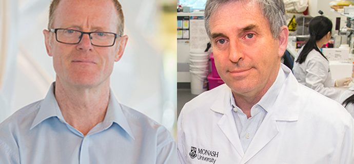
Professor Jamie Rossjohn and Professor Charles Mackay have each been ranked as a 2019 Highly Cited Researcher in the prestigious list released in November by Carivate Analytics.

Professor Rossjohn’s research focus is on using structural biology to explain pre-T- cell receptor (TCR) self-association in T-cell development, and how the TCR specifically recognises polymorphic Human Leukocyte Antigen (HLA) molecules in the context of viral immunity and aberrant T- cell reactivity. He has unearthed structural mechanisms of HLA polymorphism impacting on drug and food hypersensitivities, as well as Natural Killer cell receptor recognition. He has pioneered molecular understanding of lipid-based immunity by T cells, revealing that it can differ fundamentally from peptide-mediated adaptive immunity.

Professor Mackay has forged a new understanding of the gut microbiome and the important role it plays in immune responses including allergies and in a number of diseases including type 1 diabetes. His research into how immune responses can be manipulated using ‘medicinal foods’, as well as novel gut microbial species, is attracting both clinical and public interest, with the latest research findings moving to clinical trials.

Professor Mackay was highly cited from 2005 to 2010 under what was then the Institute for Scientific Information citation, and has remained on the Clarivate Analytics Highly Cited Researcher list since 2017.

Professor John Carroll, Director of the Monash BDI, congratulated both researchers on their achievement.

“By making the Highly Cited Researcher list two years running, Jamie and Charles have demonstrated their continued leadership in the field of immunology,” Professor Carroll said.

The Highly Cited Researcher list, now in its sixth year, determines the ‘who’s who’ of influential researchers, drawing on data and analysis to identify the world’s leading researchers who have demonstrated significant and broad influence. Seventeen researchers from Monash were recognised this year.

Original article

New grant to create biomedicine robotic art

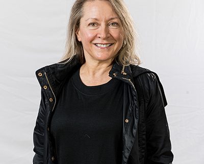
Dr Erica Tandori, artist-in-residence within the lab of ARC Laureate fellow, Professor Jamie Rossjohn, in the ARC Centre of Excellence in Advanced Molecular Imaging (Imaging CoE) at the Monash Biomedicine Discovery Institute (BDI) has been awarded a grant from Creative Victoria to explore new modelling techniques and create biomedicine art sculptures.

Over the last 18 months, Dr Tandori has developed tactile sculptures for exhibitions to explain biomedical concepts to the low vision and blind community. The newly awarded $30, 270 grant, is an opportunity for Dr Tandori to upskill and work with two techniques that she hasn’t used before – robotics and computer imaging. Using robotics and imaging, Dr Tandori will create 3D art to explain the relationship between form and function in HIV and proteins.

“As an artist, it’s amazing to be able to explore the concept by not only thinking about the picture but the actual shape. The sculptures and the exhibitions help everyone – not just the low vision community,” said Dr Tandori.

Dr Tandori will work with Swinburne’s Interaction Design Lab and Protolab to create the interactive pieces, incorporating Braille, sound and movement.  She’ll learn to use 3D technology, and explore integrating robotics, computer imaging, and organic material to explain the scientific process of protein folding and how the HIV virus infects the human body.

The sculptures give the low vision community an opportunity to hold 3D models to understand the human body, and also allow scientists and students to hold aspects of their own research.

“I can’t wait to get started! It’s such a privilege to return science to art, and art to science – just as Leonardo da Vinci once did,” Dr Tandori said.

Dr Tandori will be showcasing her current artwork at an upcoming Sensory Scientific Exhibition and Discovery Day. The half-day scientific exhibition conducted by the Monash BDI Cancer Program will be held on Friday 6 December 6. More information and register.

Dr Tandori is a legally-blind artist, academic and public speaker. She has a PhD in visual art and ophthalmology, in which she used art to articulate the processes of her own vision loss caused by juvenile macular degeneration. Since being diagnosed with the degenerative disease in her first year of art school, Dr Tandori has devoted her art-making and research to an examination of what it means to experience living with vision loss.

Original article

Out of sight – an exhibition that explains cancer to members of the low vision community

Outreach programs at universities often involve displays, exhibitions, lectures and other forms of public engagement.  Researchers within the Monash Biomedicine Discovery Institute (BDI) have developed a unique program targeted at those who have low vision or are blind.

Called the Sensory Scientific Exhibition and Discovery Day, the program has travelled nationally – most recently on show at the Lion’s Eye Institute in Perth, and was a finalist for the 2019 Eureka Prize for STEM Inclusion.

This year’s exhibition, to be held on Friday 6 December at Monash University’s Clayton campus will focus on cancer research. With tactile 3D models, 2D graphic displays, olfactory displays, large print and braille formats, the event will be specifically geared to a low vision/blind audience.

Professor Roger Daly, Head of the Monash BDI’s Department of Biochemistry and Molecular Biology and Cancer Program, is hosting the event.

“We take for granted, when talking to students, the general public and even patients, that they can see what we are talking about when describing what happens in cancer at a cellular and anatomical level,” Professor Daly said.

“For those who have low or no vision, using words is often not enough – so programs that can convert these scientific concepts into tactile and audible demonstrations are enormously valuable,” he said.

Last year, the Monash BDI’s Professor Jamie Rossjohn recruited Dr Erica Tandori, who is also legally blind, as an artist-in-residence to produce art that could explain infection and immunity to the blind and those with low vision and provide expert advice for the activities in the exhibition. This year, Dr Tandori will produce a completely new suite of tactile art and models that depict cells and cell division, tumour growth and invasion, standard and new, targeted cancer treatments and more.

Exhibition visitors will have access to 3D models of cancer cells, and immerse themselves in interactive activities and sessions on cancer research. Participants will also be able to experience Monash University’s CAVE2 facility which is a 360 degree immersive experience that will have cancer related molecules projected onto enormous surround-screens.

“We’ll have 2D and 3D models, tactile posters and interactive art. The exhibition even includes soft toys of cancer cells that can be healed when flipped inside out, so that people can experience different textures in a hands-on learning activity,” said Monash BDI’s Dr Kylie Wagstaff, who is coordinating the day.

“All of the models will be accompanied by descriptions in both large text and braille and we will have volunteers from our research community on hand to guide the participants and to explain concepts and answer questions. Each small group topic will have interactive and sensory displays suitable for all ages- we really want this to be an inspiring multi-sensory experience. We are encouraging everyone to attend, as we have activities that will appeal to all,” Dr Wagstaff said.

The Sensory Scientific Exhibition and Discovery Day will be held on Friday 6 December at Monash University, Clayton campus. Click here to find out more and to register.

Original article