Posts

Congrats Ben and team on your Nature paper

Structure of a key “trigger” of the immune response solved

An international collaboration, involving researchers from Monash University and the University of Oxford, has led to a breakthrough in our understanding of how immune responses are started. The study has just been published in Nature.

The human immune system comprises multiple important white blood cells (i.e., lymphocytes) including B cells and T cells that fight off infections and cancers. Basic discoveries leading to an understanding of how lymphocytes function have led to the development of immunotherapies and vaccines.

There are two types of T cells in humans, called αβ T-cells and γδ T cells, each of which expresses on their surfaces either an αβ T cell receptor (TCR) or a γδ TCR, respectively. In 1957, Frank Macfarlane Burnet, a famous Australian immunologist, predicted the existence of these receptors and speculated that they would “trigger” clonal lymphocyte expansions, producing enough cells to fight off infections. We now recognize that TCRs have the pivotal role of recognising molecules derived from foreign pathogens or tumours. While less is known about γδ T cells than αβ T cells, they are emerging as key players in immune defence and are becoming increasingly important for immunotherapy.

Figure. The image shows the unusual, flexible arrangement of the γδ TCR versus the αβ TCR. B-cell receptor is expressed by antibody-making B-cells.

The team determined the molecular structure of the TCR found on the surface of γδ T cells using cryogenic electron microscopy. This technically demanding project took over a decade from conception to completion and was made possible by the expertise within the Monash Ramaciotti Centre for Cryo-Electron Microscopy.

The new structure unexpectedly showed that the γδ TCR is remarkably flexible, in stark contrast to relatively rigid αβ TCRs. The work also showed that the γδ TCR is very likely the more primeval receptor and completes the initial structural analysis of Burnet’s “trigger” receptors, alongside companion paper also published in Nature.

“This flexibility is key to the ability of the γδ TCR receptor to recognise a wide array of binding partners, which underscores the unique role it plays in the human immune system”, Dr Benjamin Gully of the Monash Biomedicine Discovery Institute, co-first author of the study stated.

According to Professor Simon Davis, from Oxford University and joint senior author of the study, γδ T-cells are becoming increasingly important therapeutically.

“The new structure helps constrain theories of how TCRs trigger lymphocytes, and should be helpful, especially, for re-engineering TCRs and optimising their use in the clinic” he said.

The authors would like to acknowledge support from the Australian Research Council Discovery Program which made this basic research project possible.

Read the full paper in Nature: Structure of a fully assembled γδ T-cell antigen receptor.

DOI: 10.1038/s41586-024-07920-0

Some of the Monash University co-authors on the Nature publication (left to right): Liam Rashleigh, Michael Rice, Dr Benjamin Gully, Dilshan Gunasinghe, Professor Jamie Rossjohn, Hari Venugopal.


Original article

Monash researchers make fundamental advance in understanding T cell immunity

Monash University researchers have provided a fundamental advance regarding how T cells become activated when encountering pathogens such as viruses.

The recent study published in Science, co-led by Professor Nicole La GrutaProfessor Jamie Rossjohn and Professor Stephanie Gras with first author Dr Pirooz Zareie from the Monash Biomedicine Discovery Institute, have found that T Cells need to recognise pathogens in a particular orientation in order to receive a strong activating signal.

T cells play a key role in the immune system by eliminating invading pathogens, such as viruses, and it is crucial to understand the factors that determine how and why T cells become activated after recognizing these pathogens.

T cells express on their surface a T cell receptor (TCR) that recognizes and binds to virus fragments (antigens) presented by infected cells.  This recognition event can lead to T cell activation and killing of infected cells.

“The central issue is that there are millions of different T cell receptors (TCRs) in the human body, and a vast array of viruses, making it difficult to understand the rules around how T cell receptor recognition of a virus drives T cell activation. Indeed, it is a problem that has remained contentious for over 25 years” says Professor La Gruta.

“Our study has shown that the orientation in which the T cell receptor binds is a primary factor determining whether the T cell receives an activating signal,” Professor La Gruta said.

“This is an advance in our fundamental understanding of how a T cell needs to ‘see’ pathogenic antigens in order to be activated,” she said. “It has clarified a critical mechanism essential for effective T cell immunity. It is also relevant to the ongoing development of immunotherapies that aim to boost the activation of T cells.”

Dr Pirooz Zareie stated: “a combination of technologies, including super-resolution microscopy, X-ray crystallography at the Australian Synchrotron, biochemical assays and using in vitro and in vivo experimental models from a variety of labs led to the findings.”

The study represented a cross-disciplinary collaboration between researchers from the University of Utah, National University of Singapore, University of New South Wales and Monash University.

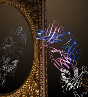
TCR-pMHC recognition - through the looking glass. The image shows a brightly colored canonical interaction between TCR and pMHC which is conducive to signal transduction. The faded mirror image shows a reversed TCR-pMHC interaction which is unable to support signal transduction and thus T cell activation. (Created by Dr. Erica Tandori (Rossjohn lab))

TCR-pMHC recognition – through the looking glass. The image shows a brightly colored canonical interaction between TCR and pMHC which is conducive to signal transduction. The faded mirror image shows a reversed TCR-pMHC interaction which is unable to support signal transduction and thus T cell activation. (Created by Dr. Erica Tandori, Rossjohn lab)

Read the full paper in Science titled: Canonical T cell Receptor Docking on peptide-MHC is essential for T cell signaling.DOI: 10.1126/science.abe9124

Original article

Also see ARC Research Highlights